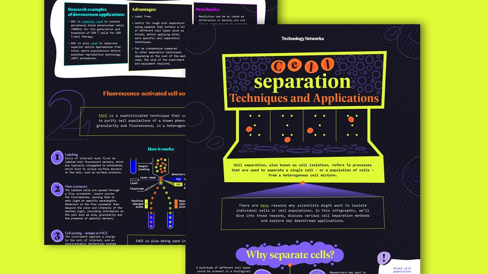
Cell Separation: Techniques and Applications
Infographic
Published: May 30, 2024
|
Molly Campbell


Senior Science Writer
Molly Campbell is a senior science writer at Technology Networks. She holds a first-class honors degree in neuroscience. In 2021 Molly was shortlisted for the Women in Journalism Georgina Henry Award.
Learn about our editorial policies

Credit: Technology Networks
Cell separation refers to processes that are used to separate a single cell – or a population of cells – from a heterogenous cell mixture. Cell separation techniques are important across all major fields of modern biology, from stem cell research to drug discovery.
Download this infographic to learn more about:
- Why scientists might want to isolate individual cells or cell populations
- Different techniques – traditional and emerging – for cell separation
- Key downstream applications of cell separation techniques
1
3
4
2
separation
Techniques and Applications
C e l l
1
2
3
“ “
1 2 3
There are many reasons why scientists might want to isolate
individual cells or cell populations. In this infographic, we’ll
dive into those reasons, discuss various cell separation methods
and explore key downstream applications.
Dr. S. H. Seal is credited with developing
the first cell separation method in
using a sieve to separate large tumor cells from smaller blood
cells. At the time, he expressed that the sieve “leaves much to
be desired” but offers a simple method for the isolation of
free-floating cancer cells.
One of the most widely used
centrifugation methods for cell
separation is density gradient
centrifugation (DGC). This method
can be used to separate cells,
organelles or macromolecules
based on their density as they
travel through a density gradient
while under a centrifugal force.
Here’s a simple overview of
the technique:
FACS is a sophisticated technique that uses flow cytometry
to purify cell populations of a known phenotype based on size,
granularity and fluorescence, in a heterogenous sample of cells.
Another affinity-based method for cell isolation, MACS utilizes
magnetic separation to isolate cells based on specific cell
surface markers. Here’s a simple overview of the technique:
Over recent years, approaches to sort cells using spatial and
time-resolved data have become increasingly popular and novel
workflows continue to emerge. Collectively, these methods are
known as image-based cell sorting, IBCS, also referred to as
image-enabled cell sorting or ICS.
Combining ICS with other omics assays, such as
protein‐ (e.g., CITE‐seq, scMS), transcript‐ (e.g.,
scRNA‐seq) and genome‐centric (e.g., scATAC‐seq,
Strand‐seq) readouts, will further increase the
resolution of single‐cell profiling experiments.
– Schraivogel and Steinmetz write.
Machine learning algorithms can be combined with real-time imaging
to support automated and precise sorting of cells in IBCS.
DGC workflow for separation of cells from a whole blood sample:
• DGC is commonly used to isolate
peripheral blood mononuclear cells
(PBMCs) for the generation and
expansion of CAR T cells for CAR
T-cell therapy.
• DGC is also used to separate
superior motile spermatozoa from
total sperm populations before
assisted reproductive technology
(ART) procedures.
Labeling
Cells of interest must first be
labeled with fluorescent markers, which
are typically conjugated to antibodies,
which bind to unique surface markers
on the cell, such as surface proteins.
FSC measures the intensity of light
scattered in the forward direction
as cells pass through the laser
beam, providing information on
the cell’s size.
Larger cells scatter more light,
resulting in higher FSC signals.
SSC measures the intensity
of light scattered at a
90-degree angle to the laser.
It provides information on the
complexity of the cell. Cells
with higher SSC signals contain
more internal structures,
such as organelles.
Sample of
heterogenous cells.
Microfluidic flow
Cells move within a fluid stream and
are imaged before being sorted into
collection containers.
Microfluidic containment
Cells are isolated and captured in
droplets or stationary traps for
standard imaging and medium throughput
sorting via fluid flow.
Microarrays
Microarrays are loaded with cells,
imaged and then are isolated using
low-throughput mechanical or optical
retrieval methods.
Select cells Sort and collect
of interest
Extract
features
Individualize Image
cells
Sample
population
Direct labeling
Cell sample is incubated with
magnetic particles that are
conjugated to antibodies targeting
specific cell surface markers.
Indirect labeling
Primary antibodies that bind
to a cell surface marker are
added to the sample. Secondary
antibodies, conjugated to
magnetic nanoparticles, are
added to the sample. They bind
to the primary antibodies.
Flow cytometry
The labeled cells are passed through
a flow cytometer. Lasers excite
the fluorophores, causing them to
emit light at specific wavelengths.
Detectors in the flow cytometer then
measure the color and intensity of the
emitted light, providing information on
the cell such as size, granularity and
the presence of specific markers.
Cell sorting – unique to FACS
The instrument applies a charge
to the cell of interest, and an
electrostatic deflection system
ensures the deflection of the charged
cells into appropriate collection
tubes, enabling cell sorting.
FACS is also being used in
combination with ultra-fast
imaging techniques to enhance
the sorting process.
• FACS has been used to sort
cells when generating knock-out
and knock-in clonal populations
of human induced pluripotent
stem cells (hiPSCs) using
CRISPR-Cas9 technology.
• Schardt et al. used FACS to purify
antigen-specific memory B cells
from mice that had been immunized
with SARS-CoV-2 antigens. This
method could identify clones
that produced potentially useful
neutralizing antibodies.
• MACS offers a quick and
reliable method for collecting
human mesenchymal stem cell
exosomes at high purity for
use in cell therapy.
• Welzel et al. established that MACS
can be used as a large-scale method
to generate zebrafish neuronal cell
cultures for neuroscience research.
• IBCS has been used in combination
with CRISPR-pooled screens to
rapidly isolate cells that have
complex phenotypes.
• Label free.
• Useful for rough bulk separation
using samples that contain a lot
of different cell types (such as
blood), before applying other,
more specific cell separation
techniques.
• Can be inexpensive compared
to other separation techniques
depending on the cost of the medium
used, the size of the experiment
and equipment required.
• Single-cell precision
and high purity.
• Can separate more than one cell
population at the same time.
• Can isolate cells based on surface
marker expression and intracellular
marker expression.
• High-throughput.
• Can be used in combination
with other methods for
downstream applications.
• Relatively low cost.
• Simple to operate.
• High selectivity.
• Relatively high speed
compared to other techniques.
• Can be used in combination with
other methods, such as FACS.
A whole blood sample is diluted
and gently layered above
the centrifugation medium,
avoiding mixing.
There are a variety of different commercialized media
types that can be used for different DGC applications.
A step gradient can be used directly, or it can be
allowed to diffuse to form a continuous gradient.
The sample is passed through a magnetic field.
The magnetically labeled cells are retained in
the field, while the unlabeled cells flow through
and can be discarded (positive selection).
The sample is then centrifuged,
and each cell type travels through
the gradient until they reach a
point where their density matches
the gradient media.
Cells can then be removed
from the interphase for
downstream applications.
Plasma
Centrifuge
Mononuclear cells
Centrifugation medium
Erythrocytes
and granulocytes
• Resolution can be an issue as
differences in density are not
always large enough to separate
individual cell types.
• Potential for sample loss during
centrifugation.
• Preparing the appropriate gradient
for the centrifugation medium can
be challenging.
• Can be time-consuming
and laborious.
• Requires specialized equipment
and training.
• Varied recovery rates.
• Requires exogenous labeling.
• The cell sorting process can be slow.
• Lacks the sensitivity and
cell-specific data that can
be provided by FACS.
• Medium throughput.
• Requires exogenous labeling.
• The process of using magnets
can be harsh on fragile cells.
To meet growing demands across various research areas, there has
been a push for the development of efficient and high-throughput
cell separation methods in recent decades.
There are now many ways to separate cells from complex samples.
The techniques used by researchers will depend on factors such as:
Next, we’ll explore four of the commonly used and emerging
methods for cell separation.
The method of choice also depends on either:
Sample
size
The physical properties of the cell
Cell size, shape or density.
Purity
requirements
Cell affinity
The electric, magnetic or adhesive properties
specific to each cell type.
Downstream
applications
Cell separation, also known as cell isolation, refers to processes
that are used to separate a single cell – or a population of cells –
from a heterogenous cell mixture.
Why separate cells?
Cell separation methods
Cell separation is important across all major fields of modern biology.
Cell separation has come a long way since then.
Density gradient centrifugation (DGC)
Fluorescence-activated cell sorting (FACS)
Magnetic-activated cell sorting (MACS)
Image-based cell sorting (IBCS)
How it works
How it works
How it works
The three main strategies used in IBCS are:
Here is an example of a typical workflow:
Research examples
of downstream applications:
Research examples
of downstream applications:
Research examples
of downstream applications:
Research examples
of downstream applications:
Advantages:
Advantages:
Advantages:
Drawbacks:
Drawbacks:
Drawbacks:
A multitude of different cell types
could be present in a biological
sample, each expressing their own
unique genes, transcripts, proteins
and metabolites that affect their
function. Single-cell analysis
requires high-throughput methods
for single-cell separation.
To isolate cells from
a tumor sample.
To analyze the effects
of a drug on a specific
group of cells.
To create
cell-based therapies.
Researchers may want to
genetically modify and
expand a specific cell
type to generate
disease models.
Mixed cell
population
Isolated cell
populations
Sample
loading
Laser beam
Laser
Electrode
Positive
charged
plate
Negative
charged
plate
Detectors
FSC Size of cells
SSC Fluorescence/
granularity
1964
1 2 3 4
Cell
number
Density (g/ml)
1.060 1.070 1.080 1.090 1.100
Monocytes
Lymphocytes
Basophils
Neutrophils
Eosinophils
Erythrocytes
1 2 3
Sponsored by
!
!
!
Magnet
High-resolution microscopy enables researchers
to visualize cell morphology, fluorescence
signals, or other markers in real-time, to
identify and select target cells.
Automated tools can be used to target and pick
cells of interest based on specific criteria
such as size, shape, spatial location of
fluorescence intensity, increasing the throughput
of cell selection.
Sophisticated tools have been developed that
enable gentle transfer of cells, preserving
cell viability for downstream applications.
Sponsored by

Download This Infographic for Free!
Information you provide will be shared with the sponsors for this content. Technology Networks or its sponsors may contact you to offer you content or products based on your interest in this topic. You may opt-out at any time.

